Cookie Policy: This site uses cookies to improve your experience. You can find out more about our use of cookies in our Privacy Policy. By continuing to browse this site you agree to our use of cookies.
AdipoGen Life Sciences
anti-Rab1-GTP, mAb (rec.) (ROF7)
As low as
150
CHF
CHF 150.00
In stock
Only %1 left
AG-27B-0006-TRIAL25 µgCHF 150.00
AG-27B-0006-C100100 µgCHF 390.00


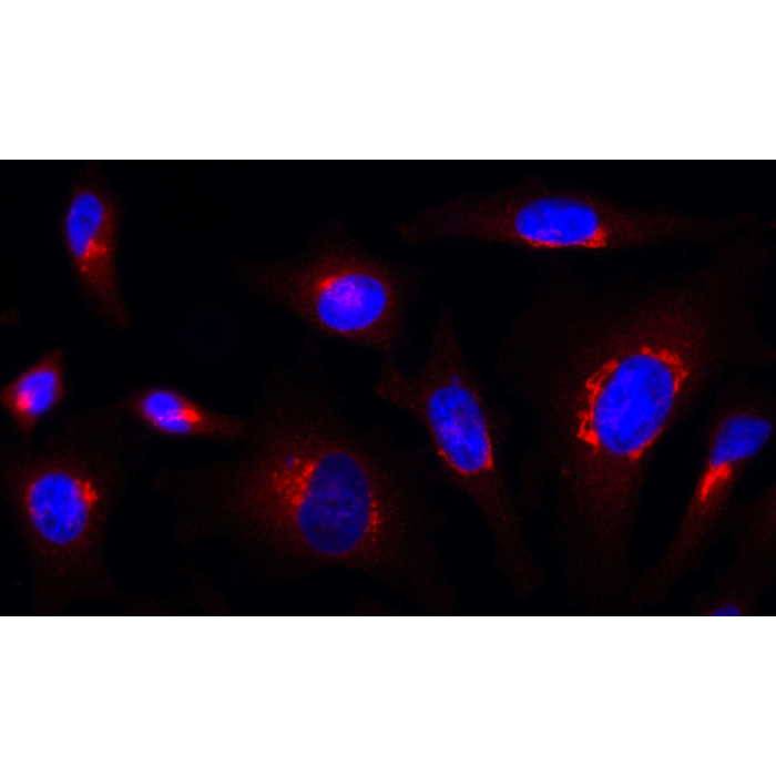
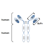
Figure 1: Schematic antibody structure.
Figure 2: Rab1-GTP is detected by immunocytochemistry using anti-Rab1-GTP, mAb (ROF7) (Prod. No. AG-27B-0006).
Method: HeLa cells are grown in standard culture conditions, fixed with paraformaldehyde (3%), permeablized in PBS+ BSA 0.2 % + Saponin 0.05 % and incubated with anti-Rab1-GTP, mAb (ROF7)(1µg /ml ) in PBS-BSA-Saponin). After incubation for 30 min at RT and several washes in PBS, cells are treated with a goat anti-human (Cy3) antibody in PBS-BSA-Saponin for 30 min at RT, washed and mounted in Moewiol. Nuclei are stained with DAPI.
Picture courtesy of Dr. Moutel, Dr. Franck Perez lab, Curie Institute, Paris.
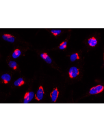
| Product Details | |
|---|---|
| Synonyms | Ras-related Protein Rab-1 |
| Product Type | Recombinant Antibody |
| Properties | |
| Clone | ROF7 |
| Isotype | Human IgG2λ |
| Source/Host | Produced without the use of animals. Purified from HEK 293 cell culture supernatant. |
| Immunogen/Antigen | Full length canine Rab1. |
| Application |
Immunocytochemistry: (1:1000) |
| Crossreactivity |
Dog Human Mouse Rat |
| Specificity |
Recognizes human, mouse, rat and dog Rab1a-GTP and Rab1b-GTP. |
| Purity | ≥95% (SDS-PAGE) |
| Purity Detail | Protein A-affinity purified. |
| Concentration | 1mg/ml |
| Formulation | Liquid. In PBS containing 10% glycerol and 0.02% sodium azide. |
| Isotype Negative Control | |
| Other Product Data |
anti-Rab1-GTP, monoclonal antibody (recombinant) (ROF7) is composed of human variable regions (VH and VL) (λ-chain) of immunoglobulin fused to the human lgG2 Fc domain. |
| Shipping and Handling | |
| Shipping | BLUE ICE |
| Short Term Storage | +4°C |
| Long Term Storage | -20°C |
| Handling Advice |
After opening, prepare aliquots and store at -20°C. Avoid freeze/thaw cycles. |
| Use/Stability |
Stable for at least 1 month after receipt when stored at +4°C. Stable for at least 1 year after receipt when stored at -20°C. |
| Documents | |
| MSDS |
 Download PDF Download PDF |
| Product Specification Sheet | |
| Datasheet |
Download PDF |
Description
Rab1 (Ypt1 in yeast) is a small GTPase that plays a well-established role in mediating ER-to-Golgi protein transport in both yeast and mammalian cells. Rab1 recruits effector proteins to budding COPII vesicles at the ER, forming cis-SNARE complexes that promote targeting to and fusion of these vesicles with the cis-Golgi. Rab1 is also involved in COPI vesicle formation and other distinct transport pathways, including ER-to-Golgi intermediate compartment (ERGIC)-to-cell periphery trafficking. Recently Rab1 has been shown to function in antimicrobial autophagy (autophagosome formation), as well as other forms of autophagy in mammalian cells in a way independent of ER-to-Golgi trafficking.
anti-Rab1-GTP, monoclonal antibody (recombinant) (ROF7) is an antibody developed by antibody phage display technology using a human naive antibody gene library. These libraries consist of scFv (single chain fragment variable) composed of VH (variable domain of the human immunoglobulin heavy chain) and VL (variable domain of the human immunoglobulin light chain) connected by a polypeptide linker. The antibody fragments are displayed on the surface of filamentous bacteriophage (M13). This scFv was selected by affinity selection on antigen in a process termed panning. Multiple rounds of panning are performed to enrich for antigen-specific scFv-phage. Monoclonal antibodies are subsequently identified by screening after each round of selection. The selected monoclonal scFv is cloned into an appropriate vector containing a Fc portion of interest and then produced in mammalian cells to generate an IgG like scFv-Fc fusion protein.
Product References
- Characterization of single chain antibody targets through yeast two hybrid: O. Vielemeyer, et al.; BMC Biotechnol. 10, 59 (2010)
- The TRAPP complex mediates secretion arrest induced by stress granule assembly: F. Zappa, et al.; EMBO J. 38, e101704 (2019)
Related Products
-
 anti-Rab6-GTP, mAb (rec.) (AA2) (ATTO488) AG-27B-0004TD
 -
 anti-Rab6-GTP, mAb (rec.) (AA2) AG-27B-0004