Cookie Policy: This site uses cookies to improve your experience. You can find out more about our use of cookies in our Privacy Policy. By continuing to browse this site you agree to our use of cookies.
AdipoGen Life Sciences
anti-HMGB1, mAb (rec.) (Giby-1-4)
As low as
150
CHF
CHF 150.00
In stock
Only %1 left
AG-27B-0002-TRIAL25 µgCHF 150.00
AG-27B-0002-C100100 µgCHF 350.00


Figure 1: Western blot analysis of human and rat HMGB1 using anti-HMGB1, mAb (rec.) (GIBY-1-4) (Prod. No. AG-27B-0002)
Different amounts of cell extracts from HEK293T cells (3μg, 5μg and 30μg) either transfected with a plasmid coding for rat HMGB1 (lanes 1, 2, 3) or non-transfected (lanes 4, 5, 6), were separated by SDS-PAGE under reducing conditions, transferred to nitrocellulose and incubated with anti-HMGB1,mAb (rec.) (GIBY-1-4) (1μg /ml). Proteins were visualized by a chemiluminescence detection system.
Different amounts of cell extracts from HEK293T cells (3μg, 5μg and 30μg) either transfected with a plasmid coding for rat HMGB1 (lanes 1, 2, 3) or non-transfected (lanes 4, 5, 6), were separated by SDS-PAGE under reducing conditions, transferred to nitrocellulose and incubated with anti-HMGB1,mAb (rec.) (GIBY-1-4) (1μg /ml). Proteins were visualized by a chemiluminescence detection system.
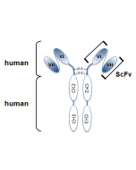
Figure 2: Schematic antibody structure.
| Product Details | |
|---|---|
| Synonyms | High Mobility Group Protein B1 |
| Product Type | Recombinant Antibody |
| Properties | |
| Clone | Giby-1-4 |
| Isotype | Human IgG2λ |
| Source/Host | Produced without the use of animals. Purified from HEK 293 cell culture supernatant. |
| Immunogen/Antigen | Human recombinant HMGB1. |
| Application |
ELISA |
| Crossreactivity |
Human Mouse Rat |
| Specificity |
Recognizes human, mouse and rat HMGB1. |
| Purity | ≥95% (SDS-PAGE) |
| Purity Detail | Protein A-affinity purified. |
| Concentration | 1mg/ml |
| Formulation | Liquid. In PBS containing 10% glycerol and 0.02% sodium azide. |
| Isotype Negative Control | |
| Other Product Data |
HMGB1, monoclonal antibody (recombinant) (Giby1-4) is composed of human variable regions (VH and VL) (λ-chain) of immunoglobulin fused to the human lgG2 Fc domain. |
| Shipping and Handling | |
| Shipping | BLUE ICE |
| Short Term Storage | +4°C |
| Long Term Storage | -20°C |
| Handling Advice |
After opening, prepare aliquots and store at -20°C. Avoid freeze/thaw cycles. |
| Use/Stability |
Stable for at least 1 month after receipt when stored at +4°C. Stable for at least 1 year after receipt when stored at -20°C. |
| Documents | |
| MSDS |
 Download PDF Download PDF |
| Product Specification Sheet | |
| Datasheet |
Download PDF |
Description
HMGB1 was originally discovered as an essential DNA-binding protein for regulating p53, NF-κB and other important proteins. It is secreted from activated dentric cells, macrophage and nectrotic cells, and acts as a ligand for RAGE, TLR-2 and TLR-4 expressed on surrounding cells. As a result, HMGB1 activates Rac, CDC42 and NF-κB inducing localized innate immunity of damaged tissue, tissue regeneration by recruitment of stem cells and hemostasis by induction of tissue factor expression. HMGB1 is also a causative agent of various diseases as it causes localized inflammation such as arteriosclerosis, chronic rheumatoid arthritis and nephritis.
Anti-HMGB1, mAb (recombinant) (Giby-1-4) is an antibody developed by antibody phage display technology using a human naive antibody gene library. These libraries consist of scFv (single chain fragment variable) composed of VH (variable domain of the human immunoglobulin heavy chain) and VL (variable domain of the human immunoglobulin light chain) connected by a polypeptide linker. The antibody fragments are displayed on the surface of filamentous bacteriophage (M13). This scFv was selected by affinity selection on antigen in a process termed panning. Multiple rounds of panning are performed to enrich for antigen-specific scFv-phage. Monoclonal antibodies are subsequently identified by screening after each round of selection. The selected monoclonal scFv is cloned into an appropriate vector containing a Fc portion of interest and then produced in mammalian cells to generate an IgG like scFv-Fc fusion protein.
Related Products
-
 HMGB1 (rat) (rec.) (His) CHI-RR-300HMGB1
 -
HMGB1 (rat):Fc (human) (rec.) CHI-RF-311HMGB1
-
HMGB1 (human) (rec.) (His) CHI-HR-200HMGB1
-
 anti-HMGB1, mAb (rec.) (Giby-1-4) (Biotin) AG-27B-0002B