Cookie Policy: This site uses cookies to improve your experience. You can find out more about our use of cookies in our Privacy Policy. By continuing to browse this site you agree to our use of cookies.
AdipoGen Life Sciences
anti-Poly(ADP-ribose) [PAR], mAb (10H)
As low as
240
CHF
CHF 240.00
In stock
Only %1 left
AG-20T-0001-C05050 µgCHF 240.00
AG-20T-0001-C100100 µgCHF 350.00
AG-20T-0001-M0011 mgCHF 1’050.00


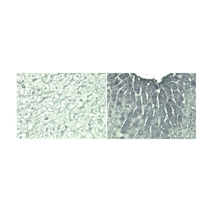
Figure 1: Immunohistochemistry of rat livers stained with anti-PAR, mAb (10H) (Prod. No. AG-20T-0001) diluted at 1:100 and treated with diethylnitrosamine (200 mg/kg) (right) or untreated (left). After treatment livers were removed and rapidly processed (10 hr later), at peak polymer induction.
Figure 2: Immunofluorescence staining of U937 cells using anti-PAR, mAb (10H) (Prod. No. AG-20T-0001). H2O2-induced PARP activation appears initially extranuclearly (10–60 minutes), while at subsequent time points nuclear PARP activation develops. Shown are images for poly(ADP-ribose), Complex I (a mitochondrial localization marker), and DAPI (a nuclear localization marker) and merged images. (see A. Brunyanszki, et al.; Mol. Pharmacol. 86, 450 (2014)).
| Product Details | |
|---|---|
| Product Type | Monoclonal Antibody |
| Properties | |
| Clone | 10H |
| Isotype | Mouse IgG3κ |
| Source/Host | Purified from concentrated hybridoma tissue culture supernatant. |
| Immunogen/Antigen | Purified poly(ADP-ribose). |
| Application |
Flow Cytometry: (see A. Kunzmann, et al.; Immun. Ageing 3, 8 (2006)) |
| Crossreactivity |
Drosophila Human Mouse Rat |
| Specificity |
Recognizes poly(ADP-ribose) synthesized by a broad range of PARPs (poly(ADP-ribose) polymerases), including human, mouse, rat or Drosophila PARP enzymes. |
| Purity | ≥95% (SDS-PAGE) |
| Purity Detail | Protein A-affinity purified. |
| Endotoxin Content | |
| Concentration | 1 mg/ml |
| Formulation | Liquid. Containing 50mM HEPES, pH 7.4, 100mM NaCl, 1% BSA and 0.02% sodium azide. |
| Isotype Negative Control | |
| Other Product Data |
Application Notes: The monoclonal antibody 10H is directed against poly(ADP-ribose) (PAR). After massive DNA damage (e.g. γ-irradiation or oxidative stress) PAR is detectable in the first 10 minutes and disappears later on. In keratinocytes the anti-PAR (10H) has been shown to detect UVB-induced apoptosis as early as 4 hours after irradiation, thus being superior to DNA laddering and the TUNEL assay. Due to the very large number of endonuclease-mediated DNA breaks in apoptosis, PARP (poly(ADP-ribose) polymerase) becomes strongly activated during the so-called execution phase. In the case of DNA damage-induced apoptosis, this represents a "second round" of PAR synthesis. PAR synthesized during apoptosis appears to be remarkably stable. PAR immunofluorescence appears at least as early during apoptosis as does the specific cleavage of PARP by caspase-3 and correlates well with other markers of apoptosis. anti-PAR (10H) was used in flow cytometry and a quantitative non-isotopic immuno-dot-blot method for the assessment of cellular poly(ADP-ribosyl)ation capacity. |
| Shipping and Handling | |
| Shipping | BLUE ICE |
| Short Term Storage | -20°C |
| Long Term Storage | -80°C |
| Handling Advice |
After opening, prepare aliquots and store at -20°C. Avoid freeze/thaw cycles. |
| Use/Stability |
Stable for at least 1 year after receipt when stored at -80°C. Stable for at least 1 week when stored at +4°C. |
| Documents | |
| MSDS |
 Download PDF Download PDF |
| Product Specification Sheet | |
| Datasheet |
Download PDF |
Description
Processes such as transcription, repair and replication that require efficient DNA recognition are dependent on modulation of chromatin structure. Chromatin relaxation is a critical event that occurs during DNA repair and is associated with the negatively charged polymer of adenosine 5'-diphosphate (ADP)-ribose (PAR). PAR is synthesized from nicotinamide adenine dinucleotide (NAD+) by the poly(ADP-ribose) polymerase protein family (PARPs), of which PARP-1 (and to a lesser extent PARP-2) respond to DNA-strand breaks. PARP-1 is selectively activated by DNA strand breaks to catalyze the addition of long branched chains of PAR to a variety of nuclear proteins, most notably PARP itself. The amount of PAR formed in living cells with DNA damage is commensurate with the extent of the damage. Under DNA damage conditions, PAR undergoes a rapid turnover, with a half-life in the range of minutes, as PAR is rapidly hydrolyzed and converted to free ADP-ribose by the enzyme poly(ADP-ribose)glycohydrolase (PARG). PAR regulates not only cell survival and cell-death programmes, but also an increasing number of other biological functions with which novel members of the PARP family have been associated. These include transcriptional regulation, cell division, intracellular trafficking, inflammation and energy metabolism.
Product References
- Monoclonal antibodies to poly(adenosine diphosphate ribose) recognize different structures: H. Kawamitsu, et al.; Biochemistry 23, 3771 (1984)
- Inhibition of poly(ADP-ribosyl)ation by overexpressing the poly(ADP-ribose) polymerase DNA-binding domain in mammalian cells: J.H. Kupper et al.; J. Biol. Chem. 265, 18721 (1990)
- trans-dominant inhibition of poly(ADP-ribosyl)ation sensitizes cells against g-irradiation and N-methyl-N'-nitro-N-nitrosoguanidine but does not limit DNA replication of a polyomavirus replicon: J.H. Kuepper, et al.; Mol. Cell. Biol. 15, 3154 (1995)
- Inactivation of the poly(ADP-ribose) polymerase gene affects oxygen radical and nitric oxide toxicity in islet cells: B. Heller, et al.; J. Biol. Chem. 270, 11176 (1995)
- Poly(ADP-ribose) synthesis: a useful parameter for identifying apoptotic cells: M. Donzelli, et al.; Histochem. J. 29, 831 (1997)
- Multiparametric staining to identify apoptotic human cells: C. Negri, et al.; Exp. Cell Res. 234, 174 (1997)
- Poly(ADP-ribose) immunostaining to detect apoptosis induced by a neurotoxic fragment of prion protein: A. Buerkle, et al.; Histochem. J. 31, 711 (1999)
- Detection of poly(ADP-ribose) synthesis in Drosophila testes upon gamma-irradiation: S. Lankenau, et al.; Chromosoma 108, 44 (1999)
- 4-Amino-1,8-naphthalimide: a novel inhibitor of poly(ADP-ribose) polymerase and radiation sensitizer: A. Schlicker, et al.; Int. J. Radiat. Biol. 75, 91 (1999)
- Reactive oxygen species participate in mdr1b mRNA and P-glycoprotein overexpression in primary rat hepatocyte cultures: C. Ziemann, et al.; Carcinogenesis 20, 407 (1999)
- Selective loss of poly(ADP-ribose) and the 85-kDa fragment of poly(ADP- ribose) polymerase in nucleoli during alkylation-induced apoptosis of HeLa cells: R. Alvarez-Gonzalez, et al.; J. Biol. Chem. 274, 32122 (1999)
- Overexpression of dominant negative PARP interferes with tumor formation of HeLa cells in nude mice: evidence for increased tumor cell apoptosis in vivo: M.A. Hans, et al.; Oncogene 18, 7010 (1999)
- Quantitative nonisotopic immuno-dot-blot method for the assessment of cellular poly(ADP-ribosyl)ation capacity: R. Pfeiffer, et al.; Anal. Biochem. 275, 118 (1999) [Immuno-Dot-Blot Detection]
- Poly(ADP-ribose) polymerase cleavage during apoptosis: when and where?: C. Soldani, et al.; Exp. Cell Res. 269, 193 (2001)
- Activation and Caspase-mediated Inhibition of PARP: A Molecular Switch between Fibroblast Necrosis and Apoptosis in Death Receptor Signaling: M. Los, et al.; Mol. Biol. Cell. 13, 978 (2002)
- Flow-cytometric assessment of cellular poly(ADP-ribosyl)ation capacity in peripheral blood lymphocytes: A. Kunzmann, et al.; Immun. Ageing 3, 8 (2006) [FACS]
- Regulation of Mitochondrial Poly(ADP-Ribose) Polymerase Activation by the b-Adrenoceptor/cAMP/Protein Kinase A Axis during Oxidative Stress: A. Brunyanszki, et al.; Mol. Pharmacol. 86, 450 (2014)
- An enzyme-linked immunosorbent assay-based system for determining the physiological level of poly(ADP-ribose) in cultured cells: C. Ida, et al.; Anal. Biochem. 494, 76 (2015) [Western Blot, HeLa]
- Tankyrase inhibition preserves osteoarthritic cartilage by coordinating cartilage matrix anabolism via effects on SOX9 PARylation: S. Kim, et al.; Nat. Comm. 10, 4898 (2019)
Related Products