Cookie Policy: This site uses cookies to improve your experience. You can find out more about our use of cookies in our Privacy Policy. By continuing to browse this site you agree to our use of cookies.
AdipoGen Life Sciences
anti-Caspase-1 (p20) (mouse), mAb (Casper-1)
As low as
480
CHF
CHF 480.00
In stock
Only %1 left
AG-20B-0042-C100100 µgCHF 480.00


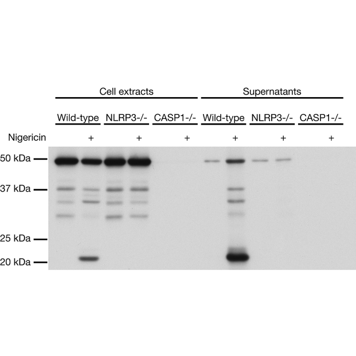
Figure 1: Mouse caspase-1 (p20) is detected by immunoblotting using anti-Caspase-1 (p20) (mouse), mAb (Casper-1) (Prod. No. AG-20B-0042). Method: Caspase-1 was analyzed by Western blot in cell extracts and supernatants of differentiated bone marrow-derived dendritic cells (BMDCs) from wild-type, NLRP3-/- and caspase-1-/- mice activated or not by 5 μM Nigericin (Prod. No. AG-CN2-0020) for 30 min. Cell extracts and supernatants were separated by SDS-PAGE under reducing conditions, transferred to nitrocellulose and incubated with anti-Caspase-1 (p20) (mouse), mAb (Casper-1) (1μg/ml). Proteins were visualized by a chemiluminescence detection system.
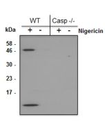
Figure 2: Immunohistochemical staining of endogenous mouse Caspase-1 in mouse spleen using anti-Caspase-1 (p20) (mouse), mAb (Casper-1) (Prod. No. AG-20B-0042). Method: Mouse spleen tissues (paraffin sections) from Caspase-1 KO (left) or WT (right) mice were stained using anti-Caspase-1 (p20) (mouse), mAb (Casper-1) (Prod. No. AG-20B-0042) (1:500) by standard immunohistochemistry (antigen retrieval performed with sodium citrate).
| Product Details | |
|---|---|
| Synonyms | Interleukin-1β Convertase; IL-1BC; Interleukin-1β-converting Enzyme; ICE |
| Product Type | Monoclonal Antibody |
| Properties | |
| Clone | Casper-1 |
| Isotype | Mouse IgG1 |
| Source/Host | Purified from concentrated hybridoma tissue culture supernatant. |
| Immunogen/Antigen | Recombinant mouse caspase-1. |
| Application |
Western Blot (see online protocol): (1μg/ml) (no need to precipitate the cell supernatant for the detection of caspase-1 (mouse) upon inflammasome activation) |
| Crossreactivity |
Mouse Rat |
| Specificity |
Recognizes endogenous full-length and activated (p20 fragment) mouse caspase-1. Described to cross-react with full-length and activated (p20 fragment) of rat caspase-1 (Lit. #30 & 34). |
| Purity | ≥95% (SDS-PAGE) |
| Purity Detail | Protein G-affinity purified. |
| Concentration | 1mg/ml |
| Formulation | Liquid. In PBS containing 10% glycerol and 0.02% sodium azide. |
| Isotype Negative Control | |
| Shipping and Handling | |
| Shipping | BLUE ICE |
| Short Term Storage | +4°C |
| Long Term Storage | -20°C |
| Handling Advice |
After opening, prepare aliquots and store at -20°C. Avoid freeze/thaw cycles. |
| Use/Stability | Stable for at least 1 year after receipt when stored at -20°C. |
| Documents | |
| Protocols |
 Download PDF Download PDF |
| MSDS |
Download PDF |
| Product Specification Sheet | |
| Datasheet |
Download PDF |
Description
Caspase-1 is the best-described inflammatory caspase. It processes the cytokines interleukin-1β (IL-1β) and IL-18 and induces pyroptotic cell death. Caspase-1 is activated by multiprotein complexes called Inflammasomes in response to numerous stimuli that are detected through distinct inflammasomes. NLRC4 responds to cytosolic flagellin, murine NLRP1b responds to anthrax lethal toxin, AIM2 responds to cytosolic DNA and NLRP3 responds to a variety of agonists including crystals.
Product References
- Measuring the inflammasome: O. Gross; Methods Mol. Biol. 844, 199 (2012)
- Inflammasome Activators Induce Interleukin-1alpha Secretion via Distinct Pathways with Differential Requirement for the Protease Function of Caspase-1: O. Gross, et al.; Immunity 36, 388 (2012)
- Omega-3 Fatty Acids Prevent Inflammation and Metabolic Disorder through Inhibition of NLRP3 Inflammasome Activation: Y. Yan, et al.; Immunity 38, 1154 (2013)
- LRRFIP2 negatively regulates NLRP3 inflammasome activation in macrophages by promoting Flightless-I-mediated caspase-1 inhibition: J. Jin, et al.; Nat. Commun. 4, 2075 (2013)
- iGLuc: a luciferase-based inflammasome and protease activity reporter: E. Bartok, et al.; Nat. Methods 10, 147 (2013)
- Receptor interacting protein kinase 2–mediated mitophagy regulates inflammasome activation during virus infection: C. Lupfer, et al.; Nat. Immunol. 14, 480 (2013)
- Activation of the NLRP3 inflammasome by IAV virulence protein PB1-F2 contributes to severe pathophysiology and disease: J.L. McAuley, et al.; PLoS Pathog. 9, e1003392 (2013)
- Caspase-1 activity affects AIM2 speck formation/stability through a negative feedback loop: C. Juruj, et al.; Front. Cell. Infect. Microbiol. 3, 14 (2013)
- Inflammasome Activation by Altered Proteostasis: J.N. Shin, et al.; J. Biol. Chem. 288, 35886 (2013)
- Immunoblotting for active caspase-1: C. Jakobs, et al.; Methods Mol. Biol. 1040, 103 (2013)
- Inflammasome activation and inhibition in primary murine bone marrow-derived cells, and assays for IL-1α, IL-1β, and caspase-1: K.s. Schneider, et al.; Methods Mol. Biol. 1040, 117 (2013)
- FADD and caspase-8 mediate priming and activation of the canonical and noncanonical Nlrp3 inflammasomes: P. Gurung, et al.; J. Immunol. 192, 1835 (2014)
- Salmonella exploits NLRP12-dependent innate immune signaling to suppress host defenses during infection: Md. H. Zaki, et al.; PNAS 111, 385 (2014)
- XIAP Restricts TNF- and RIP3-Dependent Cell Death and Inflammasome Activation: M. Yabal, et al.; Cell Rep. 7, 1796 (2014)
- The adaptor ASC has extracellular and 'prionoid' activities that propagate inflammation: B.S. Franklin, et al.; Nat. Immunol. 15, 727 (2014)
- The NLRP3 inflammasome is released as a particulate danger signal that amplifies the inflammatory response: A. Baroja-Mazo, et al.; Nat. Immunol. 15, 738 (2014)
- Caspase-1 activation by NLRP3 inflammasome dampens IL-33-dependent house dust mite-induced allergic lung inflammation: F. Madouri, et al.; J. Mol. Cell Biol. 7, 351 (2015)
- ZBP1/DAI is an innate sensor of influenza virus triggering the NLRP3 inflammasome and programmed cell death pathways: T. Kuriakose, et al.; Sci. Immunol. 1, aag2045 (2016)
- Assessing Caspase-1 Activation: B. Guey & V. Petrilli; Methods Mol. Biol. 1417, 197 (2016)
- IL-1beta and caspase-1 drive autoinflammatory disease independently of IL-1alpha or caspase-8 in a mouse model of familial mediterranean fever: D. Sharma, et al.; Am. J. Pathol. 187, 236 (2017)
- The DNA inflammasome in human myeloid cells is initiated by a STING-cell death program upstream of NLRP3: M.M. Gaidt, et al.; Cell 171, 1110 (2017)
- Inhibition of Dpp8/9 Activates the Nlrp1b Inflammasome: M.C. Okondo, et al.; Cell Chem. Biol. 25, 1 (2018)
- Complement Mediated Activation of the NLRP3 Inflammasome and its Inhibition by AAV Mediated Delivery of CD59 in a Model of Uveitis: B. Kumar, et al.; Mol. Ther. 26, 1568 (2018)
- IRF8 Regulates Transcription of Naips for NLRC4 Inflammasome Activation: R. Karki, et al.; Cell 173, 1 (2018)
- Caspase-1 self-cleavage is an intrinsic mechanism to terminate inflammasome activity: D. Boucher, et al.; J. Exp. Med. 215, 827 (2018)
- N-terminal degradation activates the NLRP1B inflammasome: A.J. Chui, et al.; Science 364, 82 (2019)
- Functional degradation: A mechanism of NLRP1 inflammasome activation by diverse pathogen enzymes: Science 364, eaau1330 (2019)
- NLRP3 inflammasome activation drives tau pathology: C. Ising, et al.; Nature 575, 669 (2019)
- Caspase-6 Is a Key Regulator of Innate Immunity, Inflammasome Activation, and Host Defense: M. Zheng, et al.; Cell 181, 674 (2020)
- Melatonin alleviates intervertebral disc degeneration by disrupting the IL-1β/NF-κB-NLRP3 inflammasome positive feedback loop: F. Chen, et al.; Bone Res. 8, 10 (2020) (Rat Samples)
- STING regulates metabolic reprogramming in macrophages via HIF-1α during Brucella infection: M.T.R. Gomes, et al.; PLoS Pathog 17, e1009597 (2021)
- DDX3X coordinates host defense against influenza virus by activating the NLRP3 inflammasome and type I interferon response: S. Kesavardhana, et al.; J. Biol. Chem. 296, 100579 (2021)
- Post-injury immunosuppression and secondary infections are caused by an AIM2 inflammasome-driven signaling cascade: S. Roth, et al.; Immunity 54, 648 (2021)
- Chronic clomipramine treatment increases hippocampal volume in rats exposed to chronic unpredictable mild stress: S. Zhang, et al.; Nature Transl. Psychiatry 12, 245 (2022) (Rat Samples)
- Oxidized DNA fragments exit mitochondria via mPTP- and VDAC-dependent channels to activate NLRP3 inflammasome and interferon signaling: H. Xian, et al.; Immunity 55, 1370 (2022)
- ZBP1-dependent inflammatory cell death, PANoptosis, and cytokine storm disrupt IFN therapeutic efficacy during coronavirus infection: R. Karki, et al.; Sci. Immunol. 7, eabo6294 (2022)
- Acute suppression of mitochondrial ATP production prevents apoptosis and provides an essential signal for NLRP3 inflammasome activation: B.S. Saller, et al.; Immunity 58, 90 (2025)
- Inflammasome signaling in astrocytes modulates hippocampal plasticity: K.E. Zengeler, et al.; Immunity 58, 1519 (2025)
- The actin and microtubule network regulator WHAMM is identified as a key kidney disease risk gene: D. Mukhi, et al.; Cell Rep. 44, 115462 (2025)
- Type A cholesterol-dependent cytolysins translocate to the trans-Golgi network for NLRP3 inflammasome activation: N. Xiao, et al.; Nat. Immunol. (Epub ahead of print) (2025)
Related Products
-
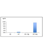
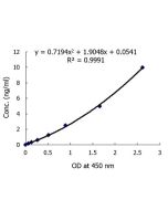 Caspase-1 (mouse) ELISA Kit AG-45B-0002
 -
 MCC950 . sodium salt AG-CR1-3615
 -
 Dapansutrile AG-CR1-3535