Cookie Policy: This site uses cookies to improve your experience. You can find out more about our use of cookies in our Privacy Policy. By continuing to browse this site you agree to our use of cookies.
AdipoGen Life Sciences
anti-LTβR (mouse), mAb (4H8 WH2)
As low as
150
CHF
CHF 150.00
In stock
Only %1 left
AG-20B-0008-TRIAL25 µgCHF 150.00
AG-20B-0008-C100100 µgCHF 320.00


Specifications / Handling
| Product Details | |
|---|---|
| Synonyms | Lymphotoxin-β Receptor; Tumor Necrosis Factor Receptor 2 Related Protein; Tumor Necrosis Factor C Receptor; Tumor Necrosis Factor Receptor Superfamily Member 3; TNFRSF3 |
| Product Type | Monoclonal Antibody |
| Properties | |
| Clone | 4H8 WH2 |
| Isotype | Rat IgG2a |
| Source/Host | Purified from concentrated hybridoma tissue culture supernatant. |
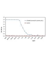
| Immunogen/Antigen | Recombinant mouse LTβR (cysteine-rich region/aa 31-221). |
| Application |
Flow Cytometry |
| Crossreactivity | Mouse |
| Specificity |
Recognizes mouse LTβR. |
| Purity | ≥95% (SDS-PAGE) |
| Concentration | 1mg/ml |
| Formulation | Liquid. In PBS containing 10% glycerol and 0.02% sodium azide. |
| Isotype Negative Control | |
| Other Product Data |
The monoclonal antibody to mouse LTβR is an agonist that can be used for the investigation of the regulation of BAFF (BlyS), chemokines and integrins using in vivo and tissue culture models, the development of NK cells and NK T cells, to study the regulation of NF-κB family of transcription factors in regulation of inflammation and homeostasis, particularly RelB NF-κB2 pathway. For use as an agonist the MAb to LTβR is added to cell cultures at 2μg/ml. For in vivo use, mice are injected intraperitoneally with 50μg of agonistic MAb to LTβR in sterile phosphate saline buffer. |
| Shipping and Handling | |
| Shipping | BLUE ICE |
| Short Term Storage | +4°C |
| Long Term Storage | -20°C |
| Handling Advice |
After opening, prepare aliquots and store at -20°C. Avoid freeze/thaw cycles. |
| Use/Stability | Stable for at least 1 year after receipt when stored at -20°C. |
| Documents | |
| MSDS |
 Download PDF Download PDF |
| Product Specification Sheet | |
| Datasheet |
Download PDF |
Scientific Background Information
Product Description
The LTβR activates two different NF-κB pathways that lead to distinct patterns of gene induction, including selected chemokines and the cytokine BAFF, which is essential for the survival of mature B lymphocytes. LTβR activates the classical NF-κB (relA/p50) pathway, like the type 1 TNF receptor (TNFR1), that regulates proinflammatory genes, like the chemokine MIP1β. However, LTβR, unlike TNFR1, also activates the processing of p100 to form RelB/p52 complexes, which activate genes involved in lymphoid organ formation and lymphocyte survival.
Product-specific References
- The lymphotoxin-beta receptor induces different patterns of gene expression via two NF-kappaB pathways: E. Dejardin, et al.; Immunity 17, 525 (2002)
- Lymphotoxin-mediated regulation of gammadelta cell differentiation by alphabeta T cell progenitors: B. Silva-Santos, et al.; Science 307, 925 (2005)
- The lymphotoxin pathway regulates Aire-independent expression of ectopic genes and chemokines in thymic stromal cells: N. Seach, et al.; J. Immunol. 180, 5384 (2008) (in vivo application)
- LTβR Signaling Induces Cytokine Expression and Up-Regulates Lymphangiogenic Factors in Lymph Node Anlagen. M.F. Vondenhoff, et al.; J. Immunol. 182, 5439 (2009)
- Quantitative Dissection and Modeling of the NF-κB p100-p105 Module Reveals Interdependent Precursor Proteolysis: Z.B. Yilmaz, et al.; Cell Rep. 9, 1756 (2014)
- Visualization of RelB expression and activation at the single-cell level during dendritic cell maturation in Relb-Venus knock-in mice: T. Seki, et al.; J. Biochem. 158, 485 (2015)
- LTβR signalling preferentially accelerates oncogenic AKT-initiated liver tumours: A.J. Scarzello, et al.; Gut 65, 1765 (2016) (in vivo application)
- Multiple roles of lymphatic vessels in peripheral lymph node development: E. Bovay, et al.; J. Exp. Med. 215, 2760 (2018)
- Molecular bases for HOIPINs-mediated inhibition of LUBAC and innate immune responses: D. Oikawa, et al.; Commun. Biol. 3, 163 (2020)
- Fibroblasts as a source of self-antigens for central immune tolerance: T. Nitta, et al.; Nat. Immunol. 21, 1172 (2020)
- Development of follicular dendritic cells in lymph nodes depends on retinoic acid-mediated signaling: J.J. Koning, et al.; Development 148, dev199713 (2021)
- Three-dimensional Imaging Reveals Immune-driven Tumor-associated High Endothelial Venules as a Key Correlate of Tumor Rejection Following Depletion of Regulatory T Cells: S. Milutinovic, et al.; Cancer Res. Commun. 2, 1641 (2022) (in vivo application)
- Simultaneous STING and lymphotoxin-β receptor activation induces B cell responses in tertiary lymphoid structures to potentiate antitumor immunity: J. Sawada, et al.; Nat. Immunol. 26, 1766 (2025) (in vivo application)
Related Products
-
 Rat IgG2b Isotype Control AG-35B-0011
 -
anti-LTβR (mouse), mAb (3C8) AG-20B-0041
-
 LTβR (mouse):Fc (human) (rec.) AG-40B-0274
 -
LTβR (human)-muIg Fusion Protein (Biotin) ANC-536-030.
-
LTβR (human)-muIg Fusion Protein ANC-536-020.
-
LTβR (human):Fc (human) (rec.) (non-lytic) CHI-HF-220LTBR
-
anti-LTβR (human), mAb (ANCLTR2/9E2) ANC-267-020.